Electron microscopy of stretch-grown axons. Scanning electron
4.5 (593) · € 33.50 · En Stock

Download scientific diagram | Electron microscopy of stretch-grown axons. Scanning electron micrographs illustrating a small fascicle composed of axons 100-250 nm in diameter (A, B). Fasciculation of axons occurs during the elongation process as smaller bundles and individual axons coalesce and adhere to one another, forming larger bundles similar to the one depicted here. Transmission electron micrograph of cross sections near the center of axon fascicles in nonstretch conditions ( C) and axons stretched to a length of 5 cm in 14 d (D), showing no change in axon cytoskeletal structures. Scale bars: A, 10 m; B, 1 m; C, D, 500 nm. from publication: Extreme Stretch Growth of Integrated Axons | Large animals can undergo enormous growth during development, suggesting that axons in nerves and white matter tracts rapidly expand as well. Because integrated axons have no growth cones to extend from, it has been postulated that mechanical forces may stimulate axon | Axons, Growth Cones and Afferent Neurons | ResearchGate, the professional network for scientists.

Magnetically-actuated microposts stimulate axon growth - ScienceDirect

Dissociated stretch-grown neurons. (A) Stretch-grown neurons following

Micromachines, Free Full-Text
IJMS, Free Full-Text

Cells, Free Full-Text

Preclinical study of peripheral nerve regeneration using nerve guidance conduits based on polyhydroxyalkanaotes - Lizarraga‐Valderrama - 2021 - Bioengineering & Translational Medicine - Wiley Online Library

Membrane Stretch Gates NMDA Receptors

Transmission Electron Microscopy and Morphometry of the CNS White Matter
Electron microscopy of stretch-grown axons. Scanning electron
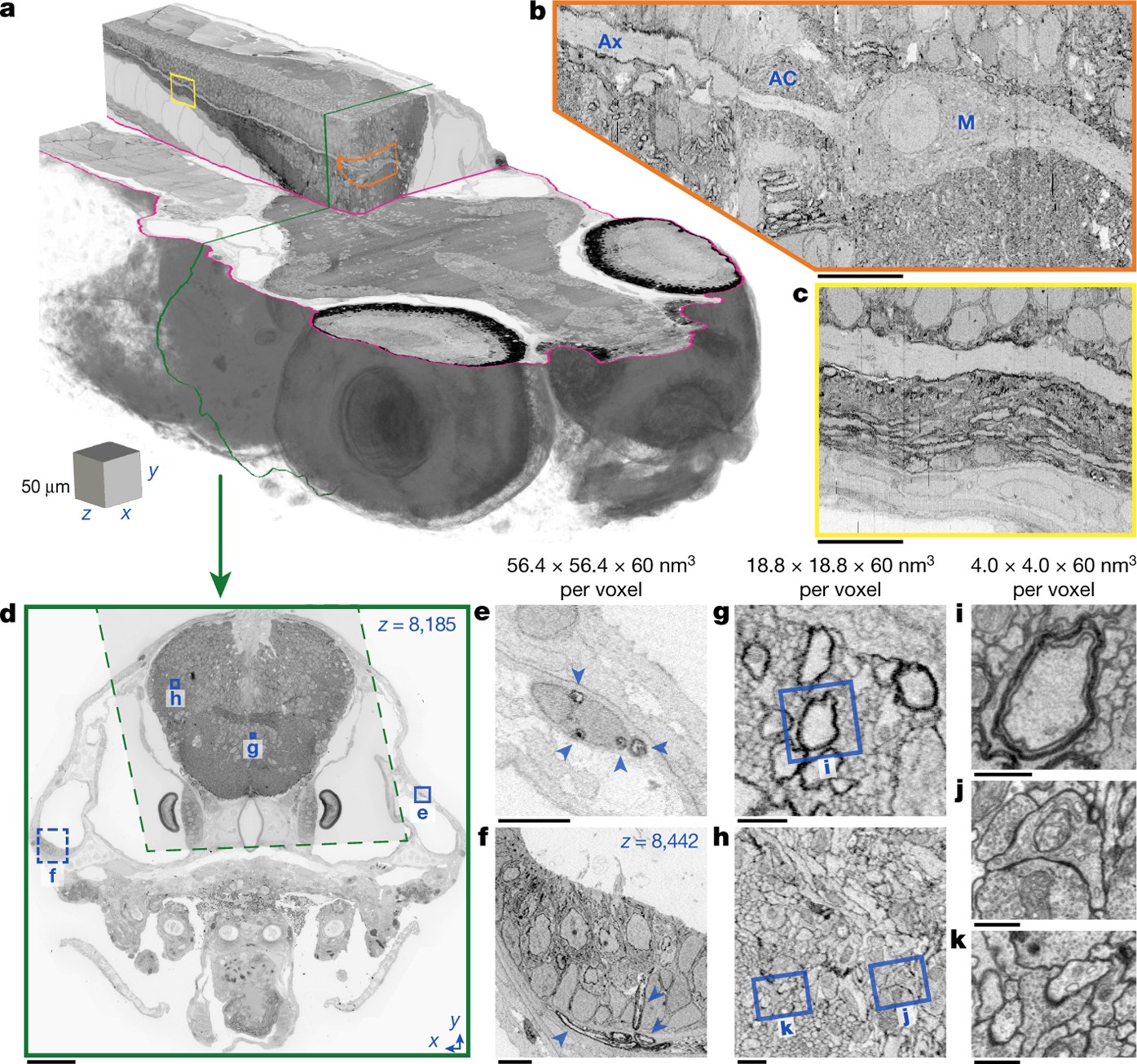
Whole-brain serial-section electron microscopy in larval zebrafish

Reconstruction of motor control circuits in adult Drosophila using automated transmission electron microscopy - ScienceDirect

Magnetically-actuated microposts stimulate axon growth - ScienceDirect