SEM (Scanning Electron Microscope) microphotographs of manganese
5 (557) · € 22.50 · En Stock

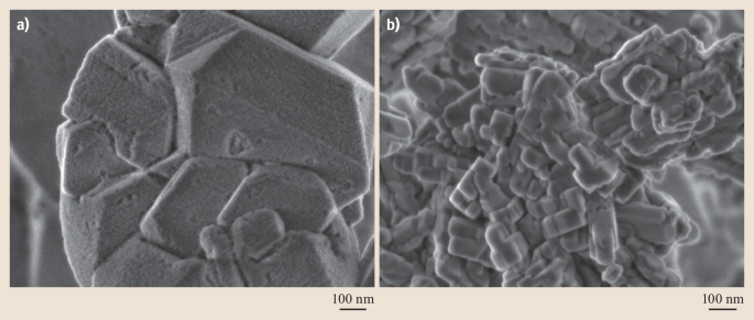
Download scientific diagram | SEM (Scanning Electron Microscope) microphotographs of manganese micronodules from the depth of 300 to 305 cm, size fraction 100-250 μm: а - micronodule with the frustules of Ethmodiscus, б - micronodule without admixture of valves of Ethmodiscus. from publication: Anomalies of rare elements in manganese micronodules from ethmodiscus oozes in the Brazil basin of the Atlantic Ocean | The composition of manganese micronodules from miopelagic clays and Ethmodiscus oozes of the central part of the Brazil Basin (station 1537, R/V Akademik Sergei Vavilov) is considered. Micronodules were recovered from >50 μm fraction of sediments from the depth intervals of | Manganese, Brazil and Atlantic Ocean | ResearchGate, the professional network for scientists.
IJERPH, Free Full-Text
Minerals, Free Full-Text
Why Use An SEM in Battery Research?

Mn-Doped Maghemite (γ-Fe2O3) from Metal–Organic Framework

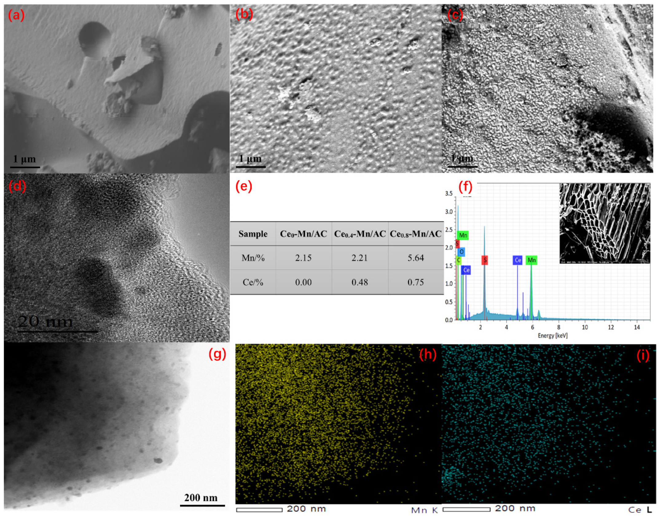
Scanning electron microscope (SEM) images with EDS elemental maps

Scanning electron microscopy (SEM) images of (a) Hercynite, (b

Electron Microscopy Techniques, Strengths, Limitations and
Scanning Electron Microscopy

Synthesis and characterization of ZnS:Mn/ZnS core/shell
Scanning Electron Microscopy Services

Photomicrographs - an overview
Magnification calibration standards for the SEM, Scanning Electron

Scanning electron microscope (SEM) micrograph of high-vanadium
ZEISS GeminiSEM Family

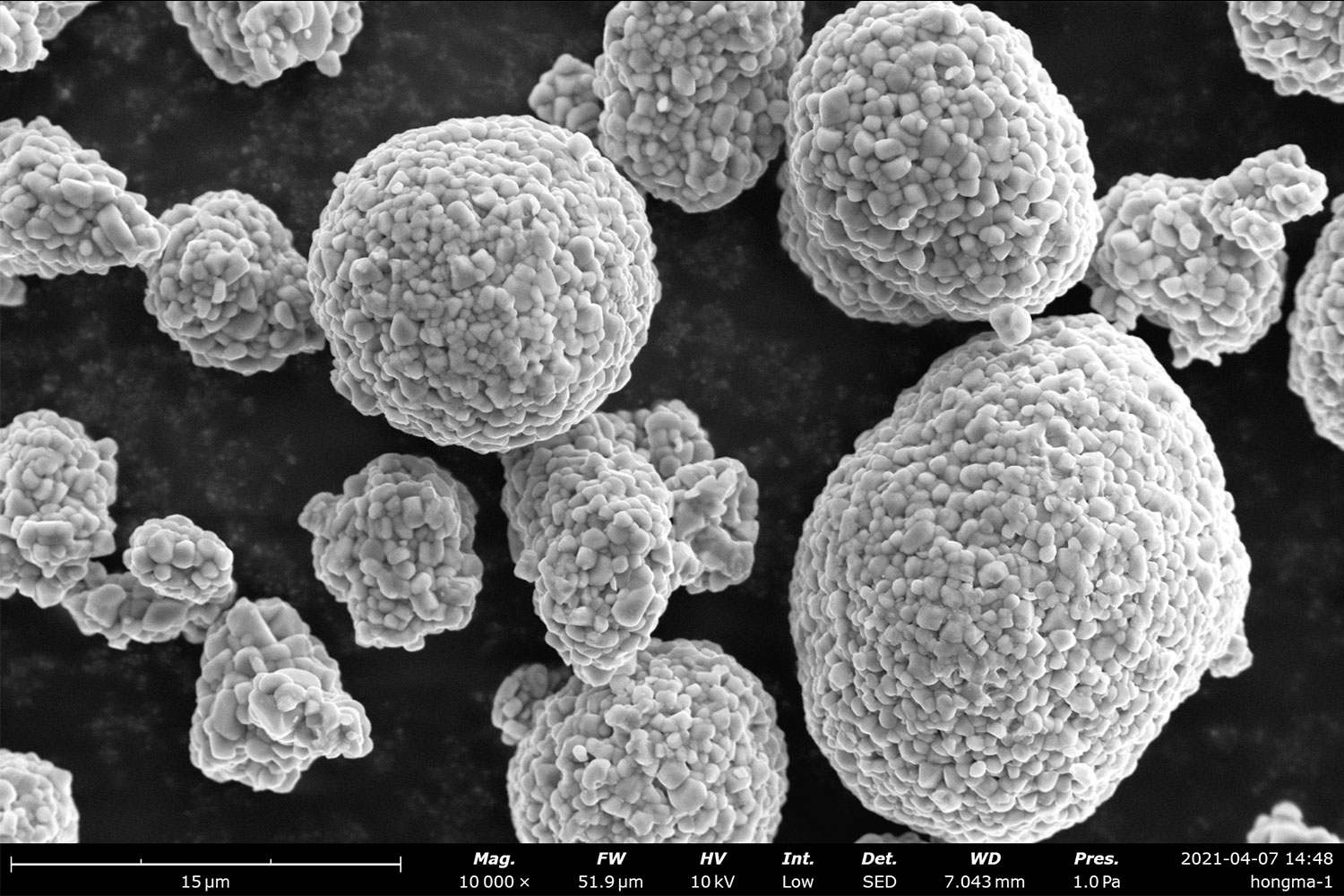
Representative scanning electron microscopy (SEM) images of (a